A Giant Parathyroid Adenoma
Abstract
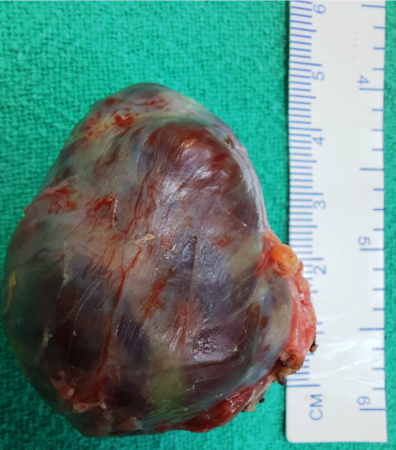
A 45-year-old female presented with a fracture of the left femur shaft after a trivial fall in the bathroom. On evaluation, the patient proved to be having severe Osteoporosis with hypercalcemia. (Sr. calcium 12.0) and markedly raised parathormone levels. (Sr. intact parathormone 428). On further biochemical investigations, the patient was diagnosed as having primary hyperparathyroidism. Examination of the neck revealed an indefinite bulge in the lower neck medial to the lower portion of the right sternocleidomastoid muscle. It was a non-tender, soft bulge which did not move with deglutition. Sestamibi scan identified an increased uptake about the lower pole of the right lobe of the thyroid with no significant uptake anywhere else in the neck. USG neck revealed a nodule approximately 4*3 cm present posterior to the lower pole of the right of the thyroid medial to Rt. common carotid artery suggestive of right inferior parathyroid adenoma. Given the concordant results of two Imaging studies, the patient was taken for focused right inferior parathyroidectomy. On exploration, a large parathyroid adenoma 5*4 cm (Fig 1) in size was present behind the lower pole of the thyroid, extending inferiorly behind the right clavicle.
Downloads

This work is licensed under a Creative Commons Attribution 4.0 International License.